
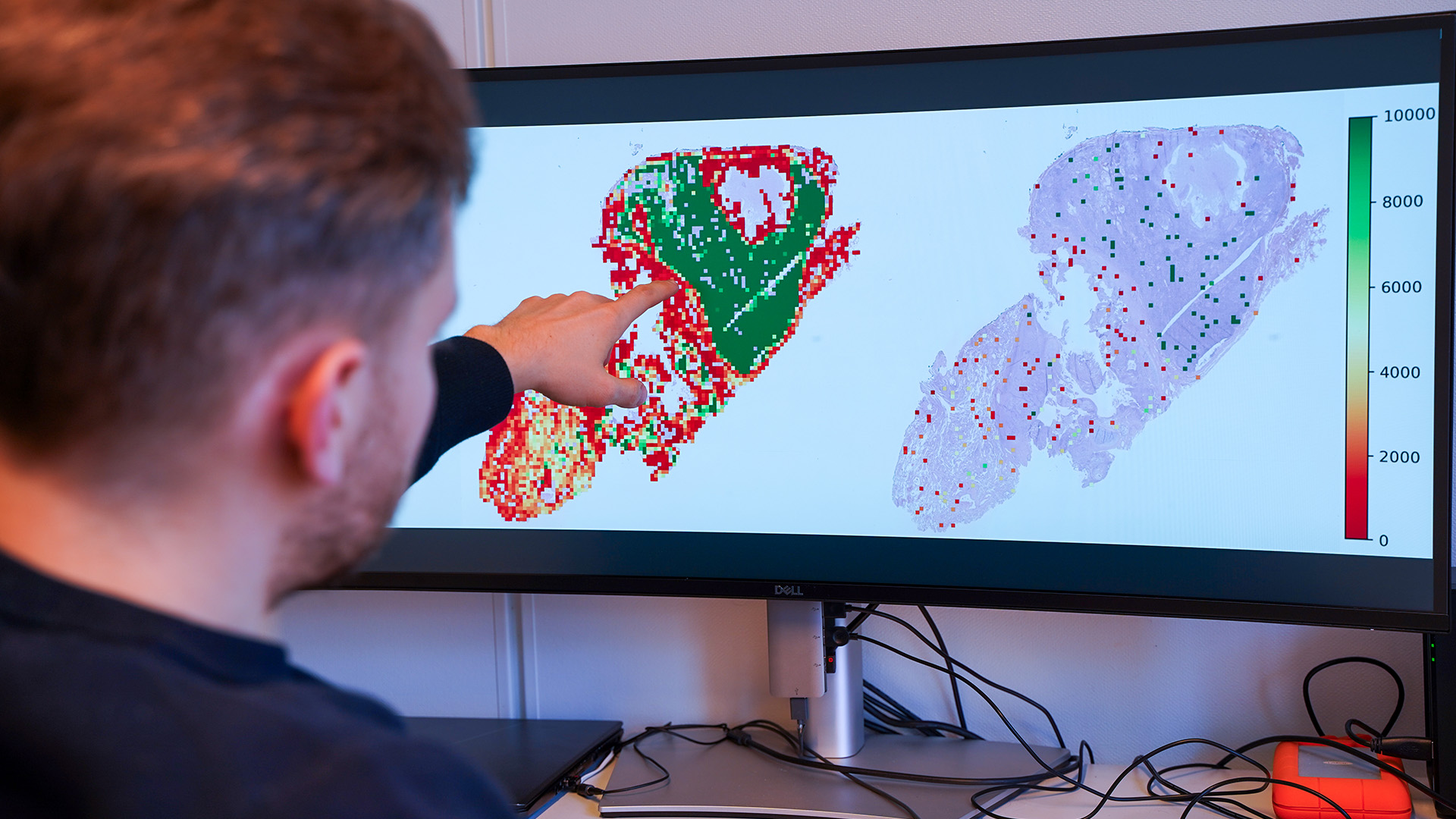
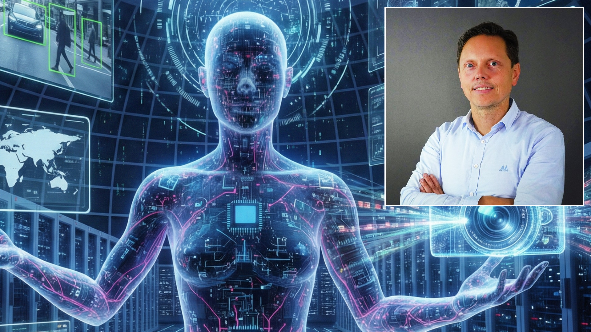
Researcher Nikita Shvetsov has developed an AI system that can contribute to less costly examinations and more personalised cancer treatment.
It can lead to less costly examinations and more personalised cancer treatment.
It can lead to less costly examinations and more personalised cancer treatment.
By Petter Bjørklund, Communications Officer at SFI Visual Intelligence
This article refers to a Visual Intelligence-authored popular science article on Science Norway.
Lung cancer is one of the most widespread and deadly types of cancer in the world. In 2023, 3,319 Norwegians were diagnosed with lung cancer.
When the immune system detects cancer inside the lungs, it responds by sending out a group of immune cells. They attack the malignant cells in the tumour.
These immune cells are called tumour-infiltrating lymphocytes (TIL). They are an integral part of the body's battle against lung cancer.
TIL cells can reveal how the cancer will develop. They can also indicate which treatment will work best.
Because of this, doctors examine tissue samples from the lungs under a microscrope. They map the TIL cells inside the tumour. The more cells they find, the better the prognosis.
The problem is that these examinations are very costly and time-consuming. The amount of TIL cells in the tissue samples may also be interpreted differently from doctor to doctor.
Researchers have now developed artificial intelligence (AI) that can make this task simpler. The technology can provide several benefits for the healthcare system – from less costly examinations to faster and more personalised cancer treatment.
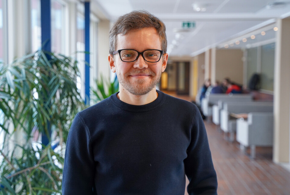
“We sought to examine how machine learning can simplify this task. Now we know that it works pretty well,” says researcher Nikita Shvetsov at UiT The Arctic University of Norway. There, he participates in a machine learning group and the AI centre SFI Visual Intelligence.
Machine learning is about enabling computers to learn something they did not know before. This is done by training them on large amounts of data. In Shvetsov's case, hundreds of digital images of lung tissue.
Parts of the dataset come from the University Hospital of Northern Norway. Researchers there are close collaborators in the researchproject.
In this way, the AI system has learnt what lungs look like – including healthy, diseased, and dead lung tissue. This allows it to filter out image sections that don't contain tissue.
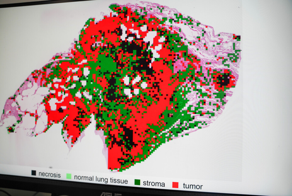
The technology falls under so-called computational pathology. It is a sub-field within digital pathology. Data-driven algorithms are used to analyse digital images of cells and tissue.
Shvetsov says this allows for more creative and automated ways of conducting these analyses.
“By using advanced algorithms and machine learning methods, the machines can find and draw out the most important sections from the images. This reduces the need for manual work and the potential risk of errors,” Shvetsov explains, adding:
“Not only does it contribute to more precise analyses, but it also enables doctors to make faster decisions that directly impact the patients.”
But digital images are often large. So large that analysing the contents of a single whole slide image can take an entire day – even with AI.
One challenge is therefore to develop systems that use fewer resources in terms of computing power, electricity, and expensive, specialised graphics processors.
Shvetsov's AI system represents a different, yet more resource-efficient way of analysing such medical images.
“A key feature of our system is that it doesn't analyse the entire whole slide image. In the first step, it selects small and random parts of the image to examine. Even though it only looks at a few sections, the system still provides a reliable overview of the number of TIL cells in the tumour,” he says.
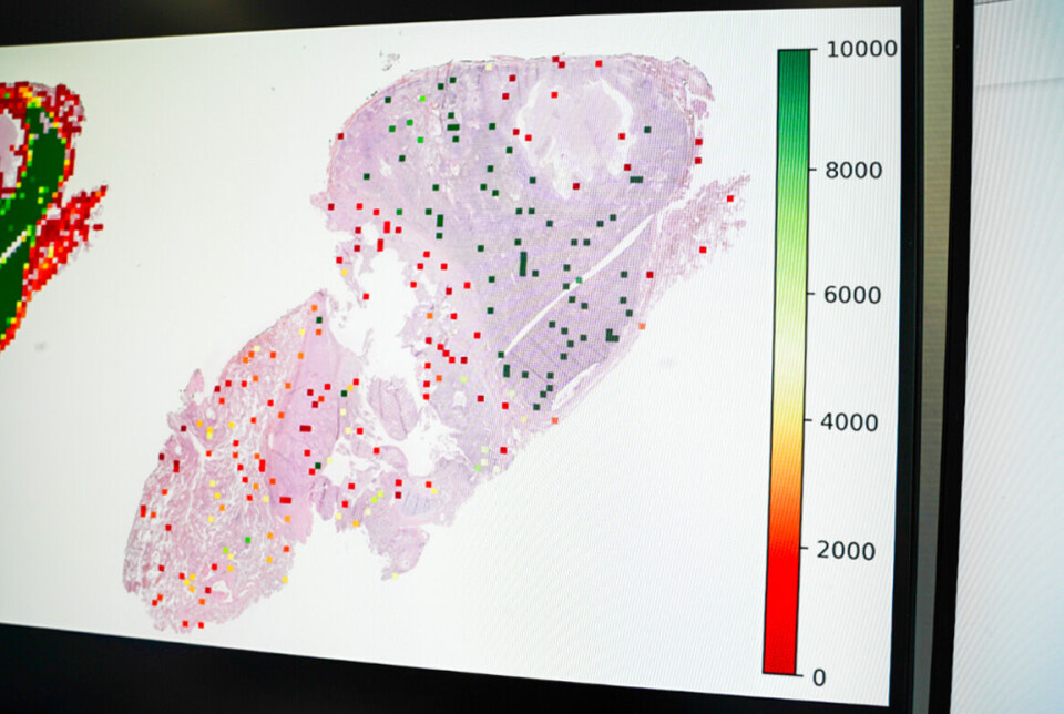
The next steps involve classifying image sections that contain TIL cells and mapping the cell quantity. This is done using an algorithm that recognises cell patterns in the image, counts different cell types, and identifies which of them are TIL cells.
The number is then converted into a score indicating the total concentration of TIL cells in different areas of the lung. Experiments from the study show that the scoring system provides an accurate indication of the patient's prognosis.
All of this is done in just a couple of minutes.
Shvetsov emphasises that the score itself is not a sufficient basis for making an accurate prognosis.
The system must be able to show how it arrived at its conclusions. This transparency is essential for establishing trust in AI as a medical tool among doctors and patients.
“In other words, the technology must be able to provide doctors with visual evidence. Our system allows them to review which cells it has identified, segmented, and classified. This allows them to verify the results and confirm that the count is actually accurate,” he says.
To check this properly, such systems must be easy for doctors to use. User-friendliness is a central focus of the research project.
“Lack of technical expertise can make healthcare professionals reluctant to adopt and use AI systems. That's why it's important that both the system and its results make sense – even for a non-expert,” Shvetsov explains.
Another goal of the project is to develop AI technology that can be adapted to other medical applications.
While the system was initially trained to map TIL cells, it can be adjusted to recognise other cell types that may also indicate the cancer prognosis.
The system can be fine-tuned to detect other biomarkers related to the body's immune response, such as PD-1 proteins.
"It can also be adapted for cancers other than lung cancer. Although this requires additional training data, integrating this into the system is quite straightforward,” he says.
While the system is still far from being a finished product, Shvetsov's research lays the groundwork for a promising medical tool.
But does that mean doctors might one day be replaced by machines that do their job?
Quite the contrary, Shvetsov replies. He stresses that AI tools are meant to assist doctors in their clinical work. No matter how advanced AI becomes, it will always be necessary to have real human beings in the loop.
“As with other AI-based tools, the goal is not to replace healthcare professionals with automated machines. Our aim is to make their lives a little easier,” he says.
Reference:
Shvetsov et al. Fast TILs—A pipeline for efficient TILs estimation in non-small cell Lung cancer, Journal of Pathology Informatics, vol. 17, 2025. DOI: 10.1016/j.jpi.2025.100437
Visual Intelligence hosted over 45 international AI researchers for the DL2026 workshop at UiT The Arctic University of Norway.
Visual Intelligence will be well represented at MICCAI 2026, one of the leading AI conferences on medical imaging and computer assisted intervention, with two accepted research papers.
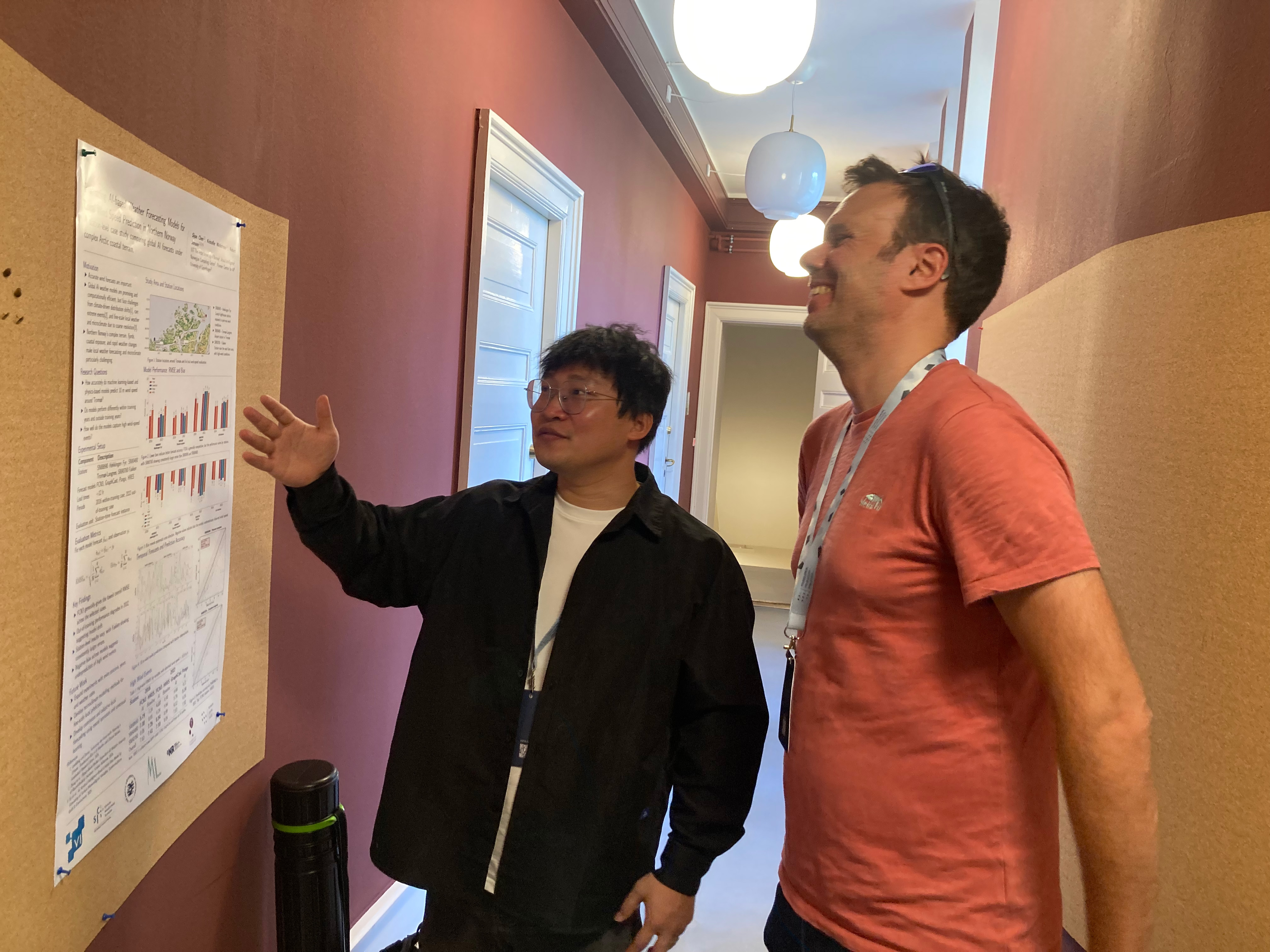
Visual Intelligence co-organized and attended the "Foundations of Arctic AI and Generative Forecasting": an inaugural workshop in the P1 Arctic AI program.
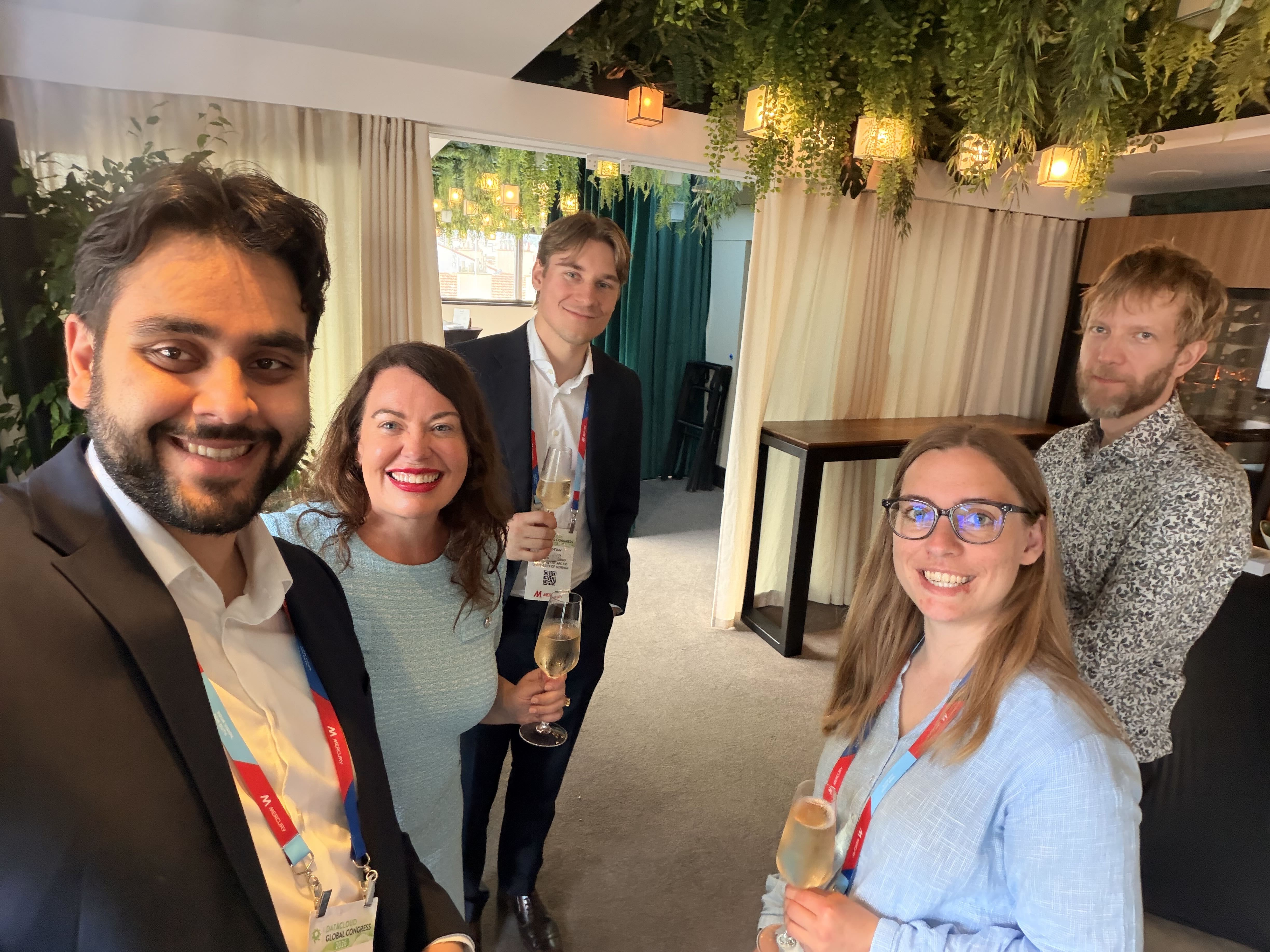
PhD Candidates Solveig Thrun and Christian Salomonsen attended the Datacloud Global Congress' Talent in Tech programme, which invited emerging talent to a unique programme designed to inspire, educate, and connect young professionals with senior leaders from global tech giants.

We warmly welcome Georgios Leontidis as a new Professor at SFI Visual Intelligence's hub at UiT The Arctic University of Norway.

Centre Director Robert Jenssen was interviewed by Norsk rikskringkasting (NRK) about the Japan-Norway AI Innovation Forum in Tokyo, Japan.

En ny AI-løsning utviklet ved UiT kan gjøre det enklere å avdekke feilrapportering og ulovlig fiske. Nå skal Norges Råfisklag teste teknologien i praktisk bruk (News article on kystogfjord.no)
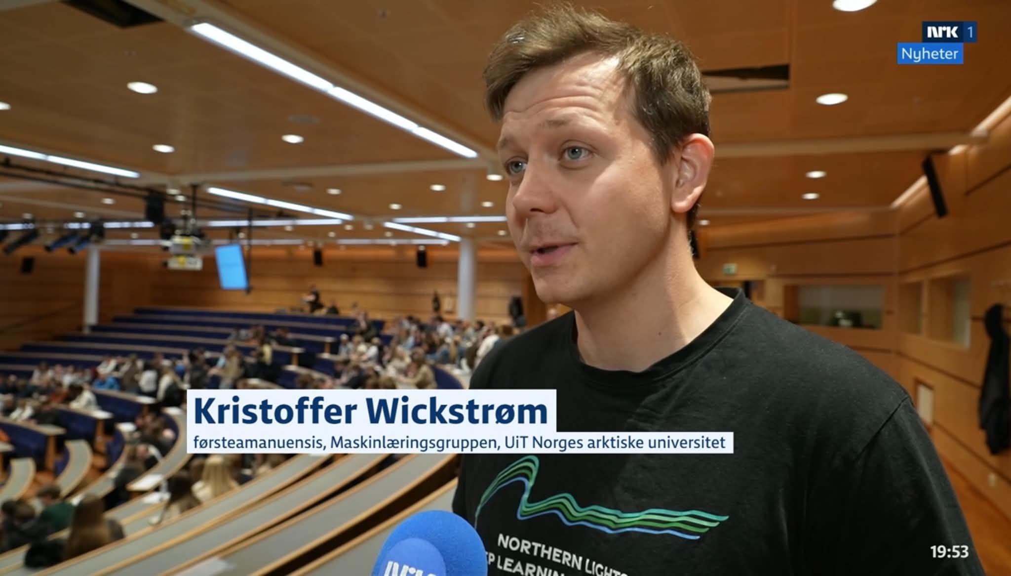
Principal Investigator Kristoffer Wickstrøm was interviewed by Norsk rikskringkasting (NRK) about the Artificial Intelligence Day at UiT- The Arctic University of Norway.
.jpg)
En ny, åpent tilgjengelig KI-modell kan endre hvordan geologer tolker seismikk. Den norske grunnmodellen lover raskere analyser, lavere terskel for innovasjon og nye måter å forstå undergrunnen på.
Centre Director Robert Jenssen was interviewed by Norsk rikskringkasting (NRK) about AI in fisheries and this year's UArctic (University of the Arctic) Congress on the Faroe Islands.
.jpg)
Professor and AI expert Robert Jenssen is attending the Japan-Norway AI Innovation Forum and Japan-Norway Research Symposium, two high-level meetings with Norwegian and Japanese government leaders, business actors and researchers.
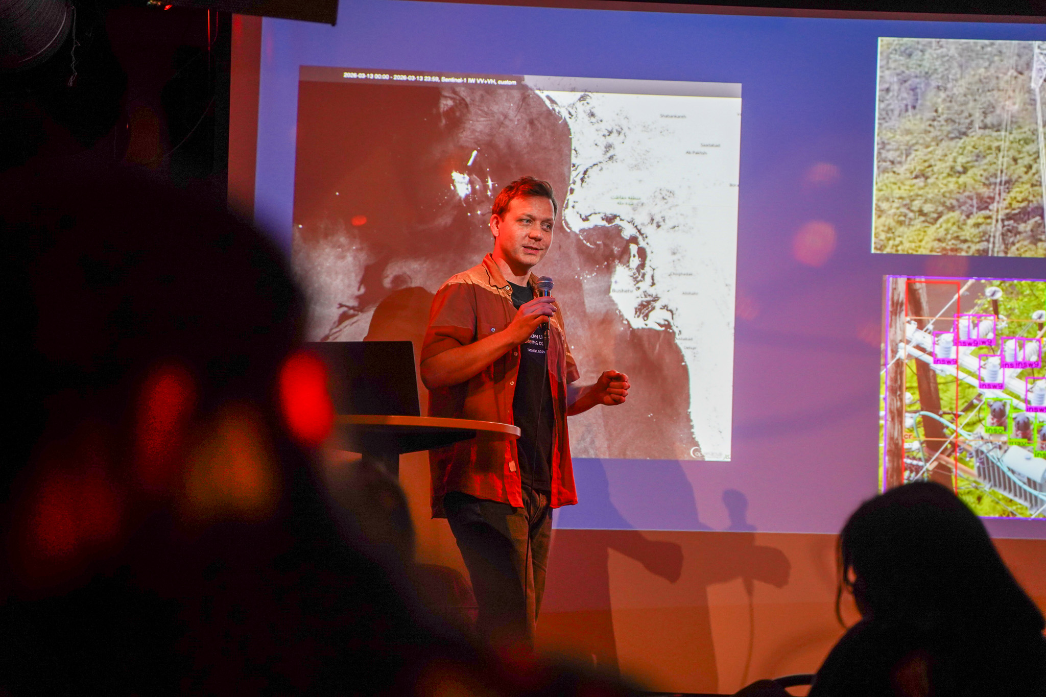
Visual Intelligence researchers Solveig Thrun and Kristoffer Wickstrøm took their research out of the lab and to Tromsø city centre as part of Pint of Science 2026.
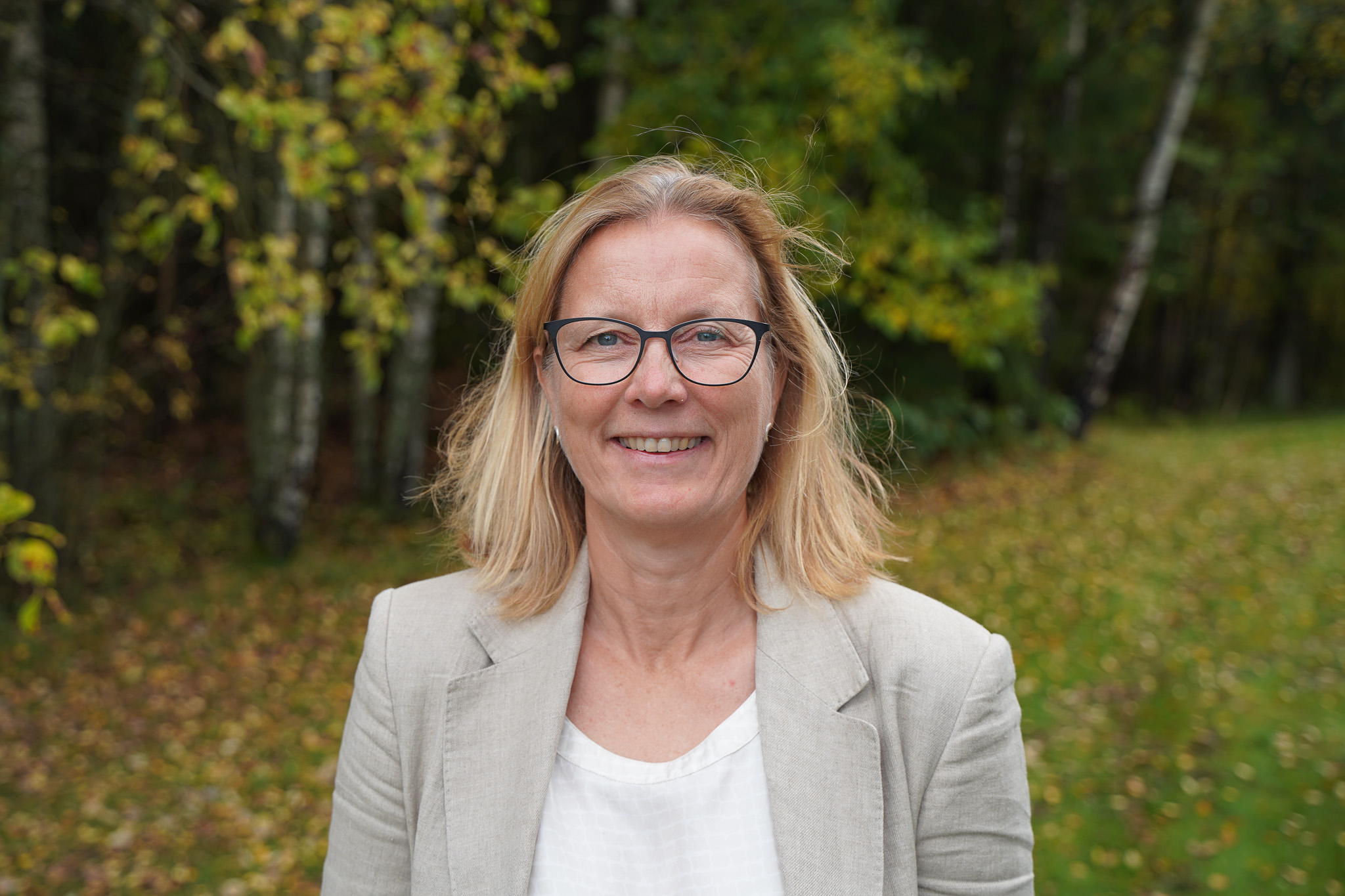
Subsurface Digital Manager and former Visual Intelligence board member Cathrine Tegnander will serve as the elected leader of Visual Intelligence's board.
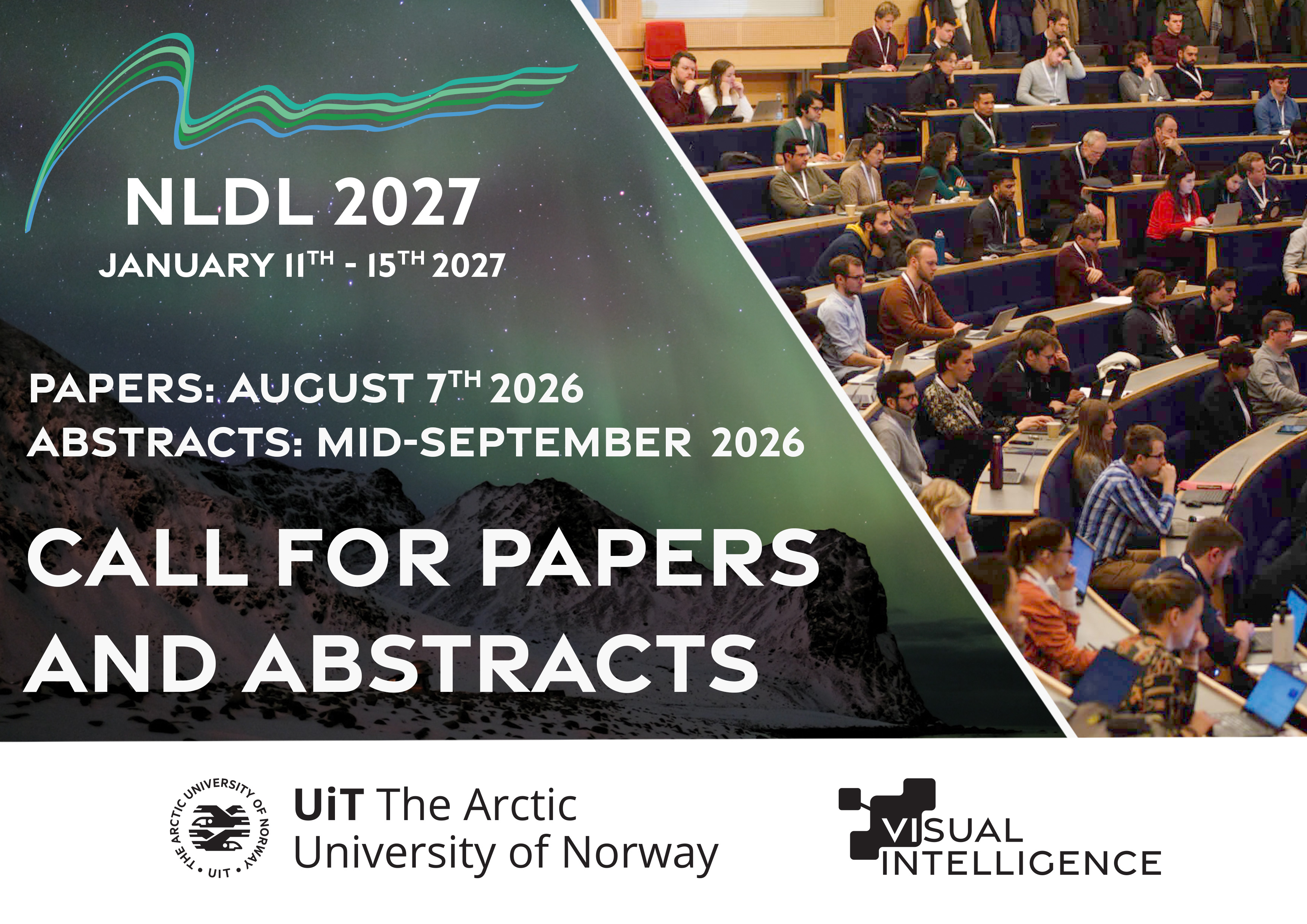
The Call for Papers and Abstracts for the Northern Lights Deep Learning (NLDL) Conference 2027 is officially announced – with submission deadlines on August 7th and Mid-September 2026 respectively.
The field of Visual Intelligence is continuously transforming. Chief Research Scientist Arnt-Børre Salberg dives deeper into the current trends in the field of visual intelligence as of early 2026.
Generativ kunstig intelligens er imponerende, men ikke alltid så nyttig til å løse industrielle problemer (Norwegian op-ed in digi.no).
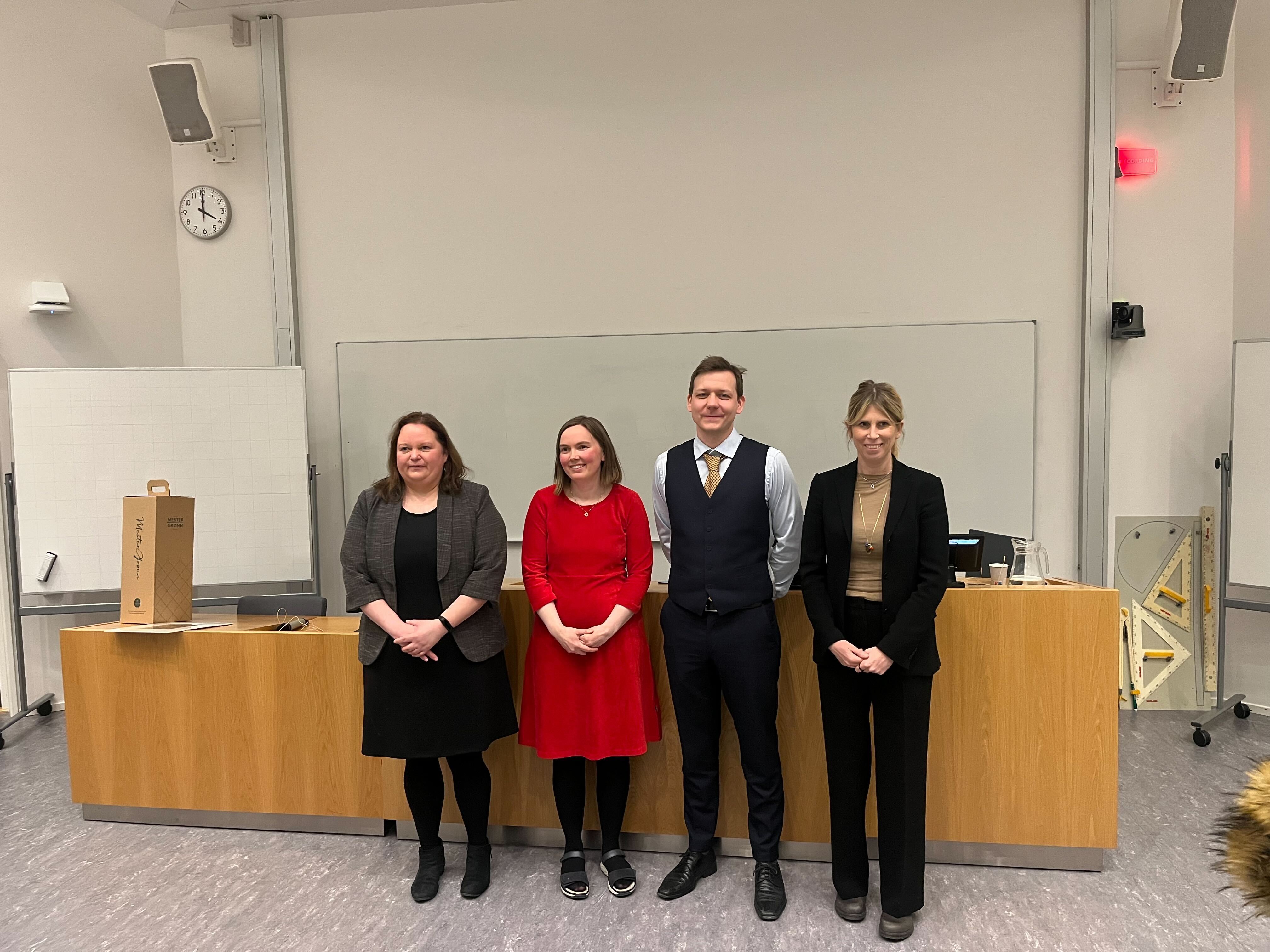
Congratulations to Marit Dagny Kristine Jenssen, who successfully defended her PhD thesis at UiT The Arctic University of Norway on April 10th

The NCS model, a seismic foundation model trained on data from the Norwegian data repository for subsurface data, is now available as an open-source model, allowing anyone to download, utilize, and further develop the model.
The Visual Intelligence Annual Report 2025, highlighting the centre's progress, activities, achieved innovations, staff, funding, and publications for 2025, is now available to read on our websites.
.jpg)
Congratulations to Centre Director Robert Jenssen, who received UiT's Research and Development Award at the university's annual celebration.