
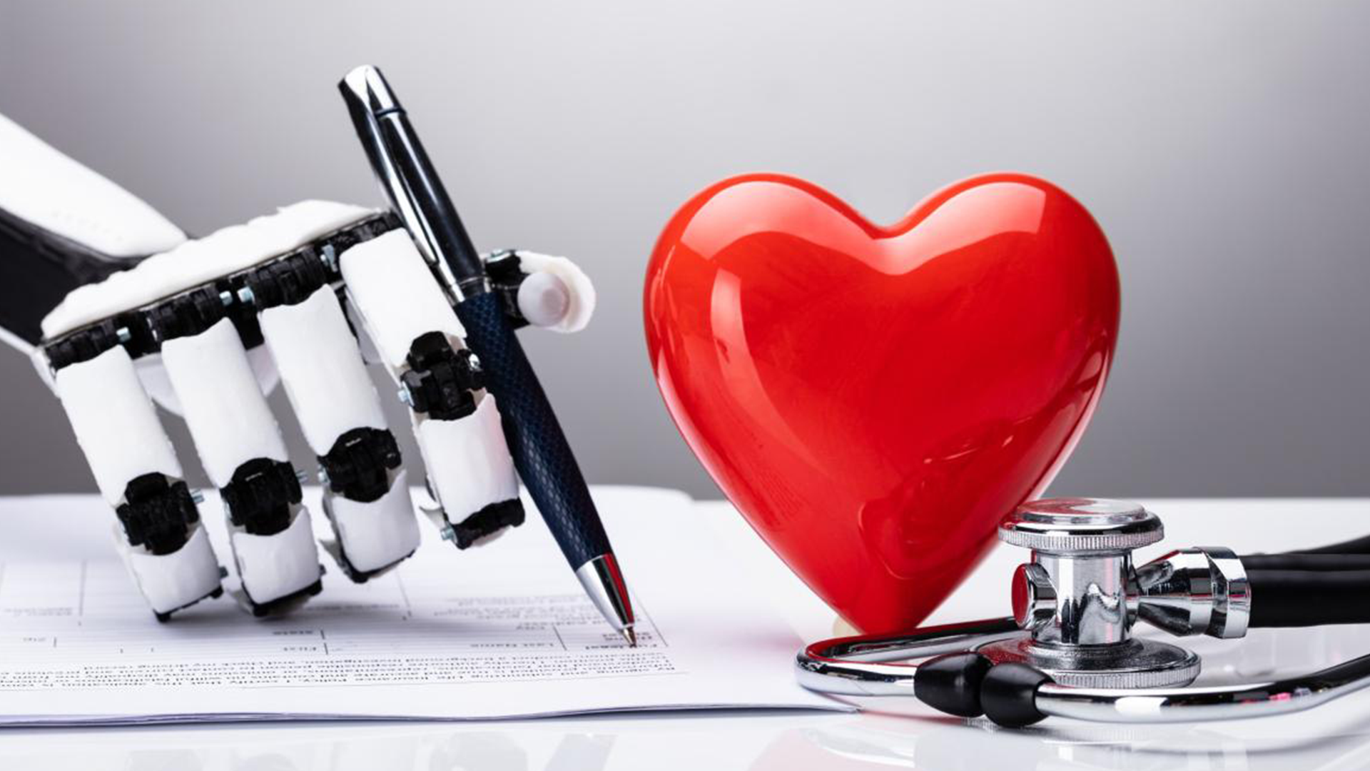
A newly developed AI system may help doctors detect and treat cardiovascular diseases more quickly.
Visual Intelligence researchers have developed an AI to automatically measure the heart's structure – both quickly and accurately. They believe it can help doctors detect and treat cardiovascular diseases faster.
UiT researchers have developed an AI to automatically measure the heart's structure – both quickly and accurately. They believe it can help doctors detect and treat cardiovascular diseases faster.
By Petter Bjørklund, Communications Officer at SFI Visual Intelligence
The news article was originally published on en.uit.no
Artificial intelligence (AI) may become a helpful tool for detecting and treating cardiovascular diseases.
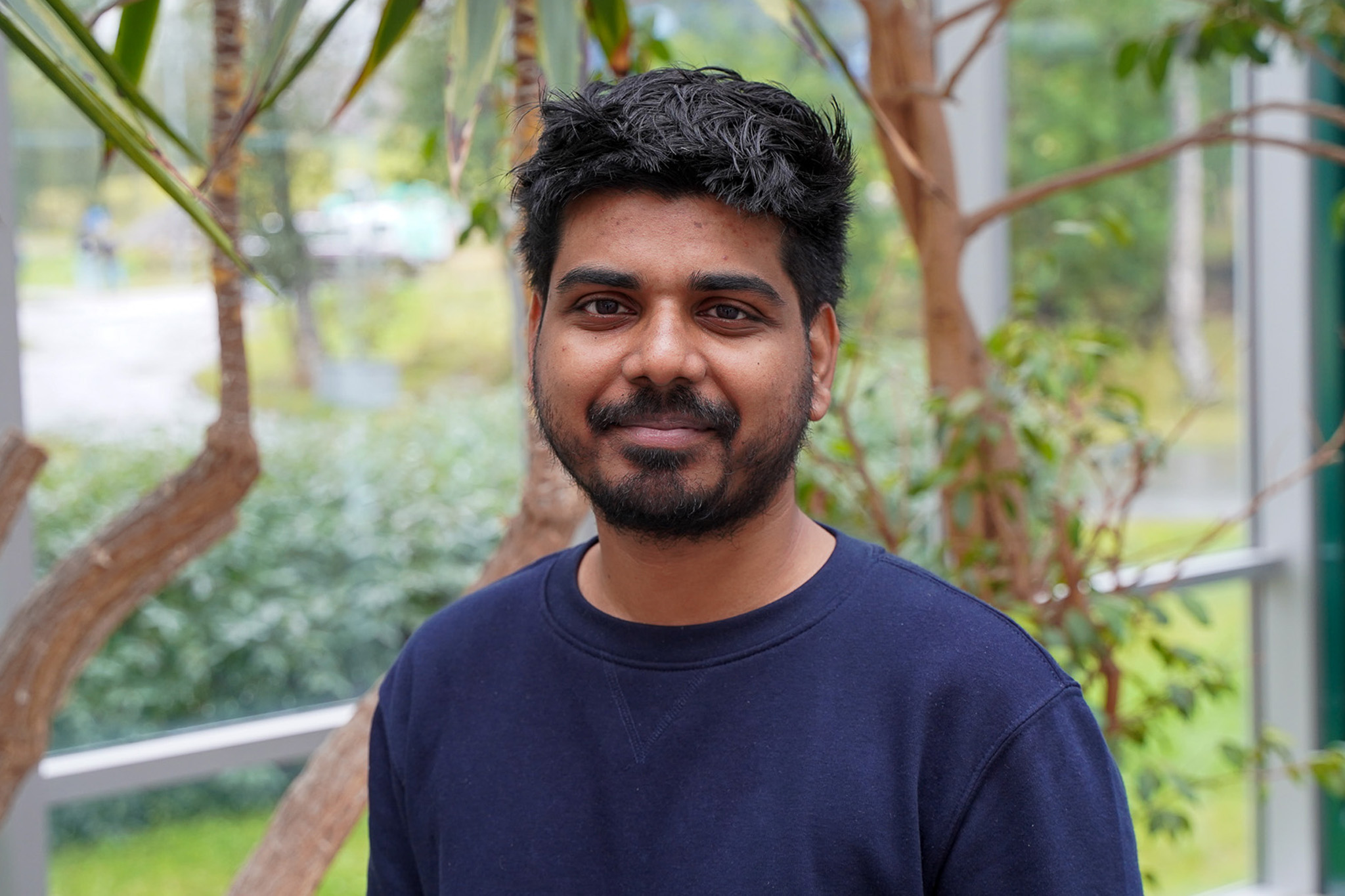
This is shown by findings from Durgesh Kumar Singh's doctoral thesis – which he defended this fall. He is a researcher at the AI centre SFI Visual Intelligence at UiT The Arctic University of Norway.
Singh examined how to use AI to measure the heart's left ventricle (LV) – one of the four heart chambers – based on heart ultrasound images. These images are obtained using an ultrasound probe which is placed on the patient's chest.
The LV is the heart's main pumping chamber and serves an important function – to pump out oxygen-rich blood to the aorta, which carries the blood to the rest of the body.
If not enough blood is pumped out, the organs won't receive enough oxygen to function properly. This may happen due to diseases or conditions which change the LV's size, thickness, or overall shape – such as high blood pressure and heart-muscle diseases.
The measurements give doctors valuable information about the LV's size and pumping capacity. This is used to detect potential signs of heart disease.
Singh's results show that his AI method can measure the heart chamber more quickly and accurately. He believes it could become a promising tool for healthcare services in and outside Norway.
"We show how deep learning can be used to measure the LV on ultrasound images more accurately and consistently by making the AI more aware of the heart's structure," Singh says.
Deep learning involves teaching machines to perform specific tasks without direct human instructions. Based on this, Singh trained an AI model using thousands of LV ultrasound images. The data set was obtained from GE HealthCare and contains images from hospitals worldwide.
Through this process, the AI taught itself what the LV looks like. It uses this knowledge to find the most important parts of the image for measuring the heart chamber.
"Think of it as an assistant which automatically lays the ruler at the correct place. The computer first finds the best measuring line by itself by outlining the heart's shape. The ruler is then used to read the size along that line," Singh explains.
Measuring the LV is usually done manually by a cardiologist. This involves examining the ultrasound image, drawing a straight line across the LV, and placing measurement points on that line. However, it is a time-consuming and delicate process, even for experienced cardiologists.
"It is careful, repetitive work that takes a lot of time. The measurements may also vary depending on the patient's heart anatomy and the person doing the examination," Singh says.
Singh's AI method can measure the LV in a matter of seconds. This provides many potential benefits – from faster answers for patients to less workload for doctors.
"Automating the most time-consuming parts means that results can be ready during the exam – speeding up diagnosis and treatment. Accurate measurements can also help track subtle heart changes more reliably over months and even years," Singh explains.
An ultrasound examination may cost hospitals and patients up to several thousand Norwegian kroner. Consistent measurements also mean fewer unnecessary exams.
"More accurate and repeatable measurements reduce do-over exams, saving patients' and the healthcare system's time and money," he says.
To examine how well it measures the heart chamber, Singh compared his method to other AI models on the market. The results show that it outperforms these models – both in terms of placing measurement points accurately and overall agreement with human experts.
Clinicians measure the LV using two types of ultrasound images: so-called B-mode and M-mode images. B-mode images represent the ultrasound waves as a two-dimensional grayscale image, while M-mode images display the heart wall's motions through a scanline.
.jpg)
Singh's method works on both types of ultrasound images, making it more useful and applicable in real clinical scenarios.
"Depending on the doctor's needs or preferences, the measurements can be displayed on either image type," Singh says.
Singh collaborated closely with GE Healthcare – one of several industrial partners in SFI Visual Intelligence. A goal of the project was to develop an AI-based algorithm which can be integrated into GE HealthCare's ultrasound scanners.
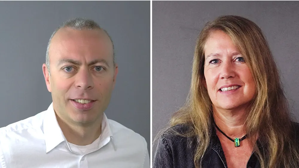
Erik Steen is a chief engineer at GE HealthCare, and says there's a need to increase the productivity of these examinations. He believes Singh's work could contribute to this.
"This need is echoed by several clients and experts worldwide. Durgesh's work can contribute to increased productivity by automating measurements needed to uncover heart diseases like thickened heart wall," Steen says.
Based on this potential, GE HealthCare plans to test Singh's AI method in a controlled and safe environment. They hope his work may eventually be incorporated into their scanners.
But it will require several rounds of testing before that happens, Steen says. This is to ensure that the method consistently measures the heart chamber correctly.
"It's important for us to check that these methods are robust and work on a large number of patients with varying image quality. We also need to document that they are just as good as or better than human experts who measure by hand," Steen explains.
Singh's findings show how useful this technology could be for detecting and treating heart diseases. But what will happen to the doctors? Will they eventually be replaced by artificially intelligent algorithms?
.jpg)
Singh assures us that this is not the case. AI is meant to assist doctors with their clinical work. The doctors will always be there to make sure that the AI does the job properly.
"It's designed to help, not replace. The technology's purpose is to remove the most tedious and time-consuming steps. The doctors are still keeping control," Singh says.
Professor Michael Kampffmeyer is Singh's main supervisor and played a key role in the doctoral project. He is impressed by Singh's work and believes it can significantly benefit both patients and doctors worldwide.
"His research is a clear example of how AI can make the healthcare system more efficient – in this case, for detecting cardiovascular diseases more quickly. We look forward to seeing how the testing at GE HealthCare will go," Kampffmeyer says.
Reference:
Durgesh Kumar Singh: Towards more accurate and label-efficient Left Ventricle Automatic Measurements. Doctoral Dissertation at UiT The Arctic University of Norway, 2025. Summary.
Visual Intelligence will be well represented at MICCAI 2026, one of the leading AI conferences on medical imaging and computer assisted intervention, with two accepted research papers.
Visual Intelligence co-organized and attended the "Foundations of Arctic AI and Generative Forecasting": an inaugural workshop in the P1 Arctic AI program.
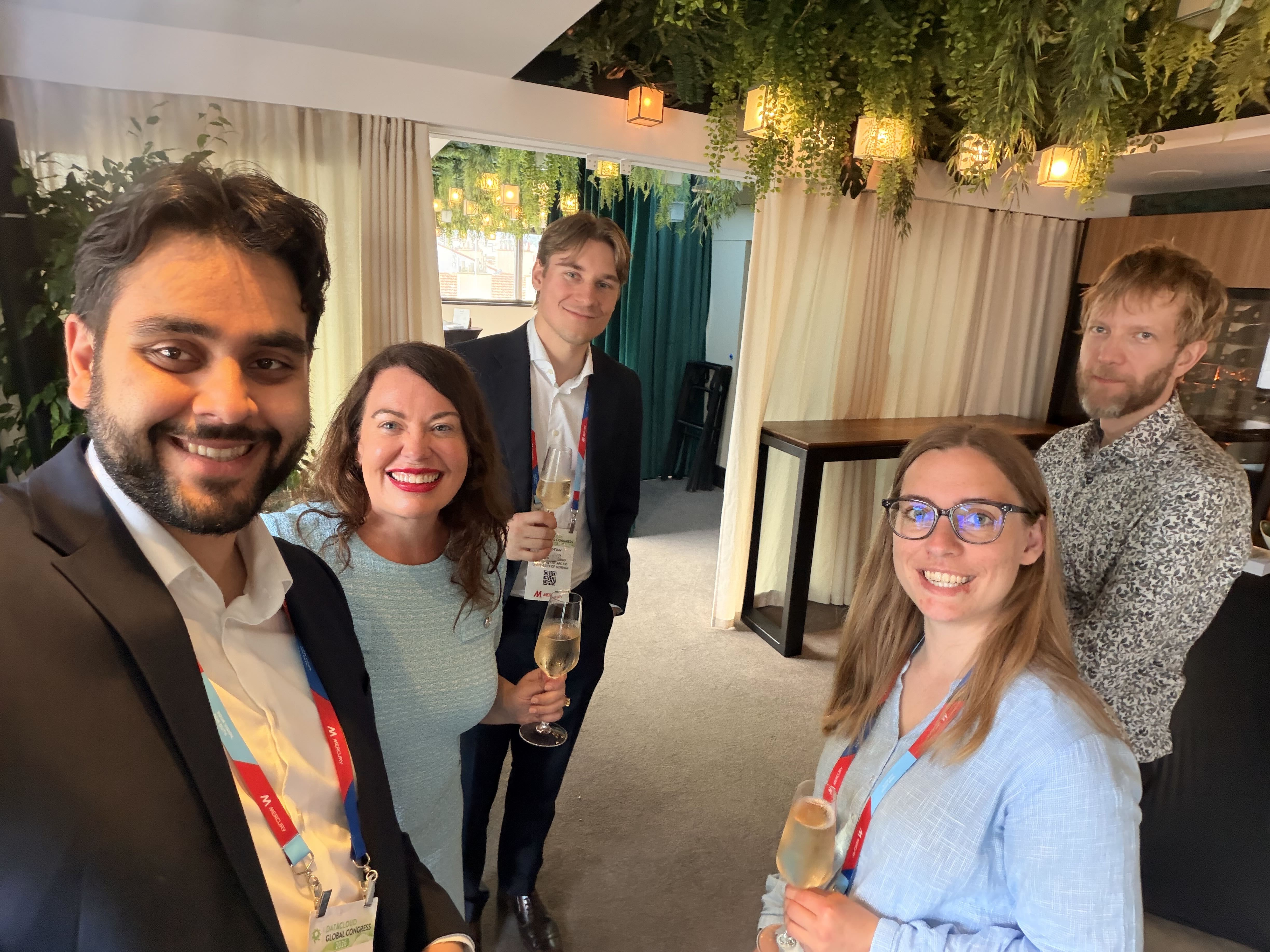
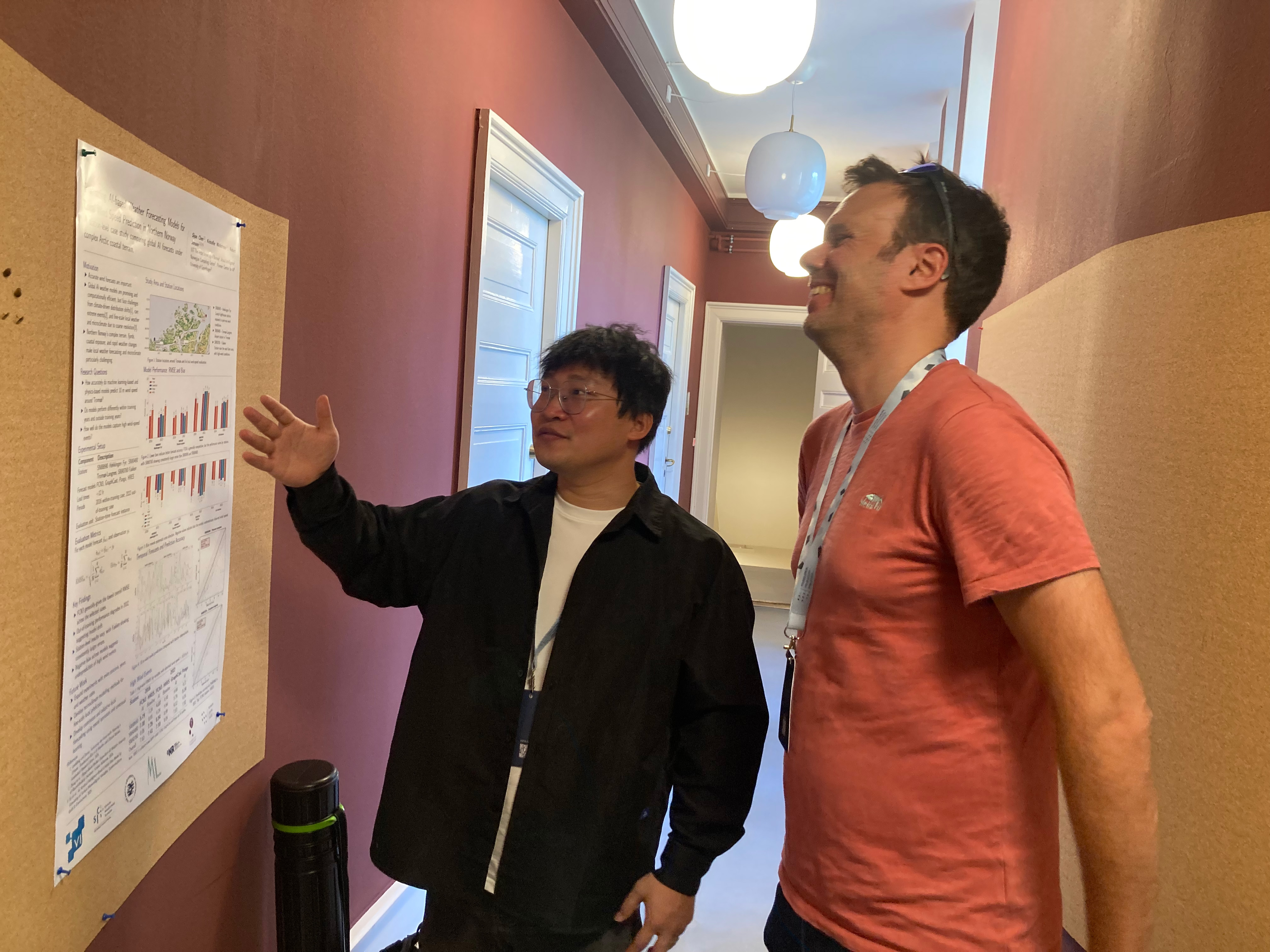
PhD Candidates Solveig Thrun and Christian Salomonsen attended the Datacloud Global Congress' Talent in Tech programme, which invited emerging talent to a unique programme designed to inspire, educate, and connect young professionals with senior leaders from global tech giants.
We warmly welcome Georgios Leontidis as a new Professor at SFI Visual Intelligence's hub at UiT The Arctic University of Norway.
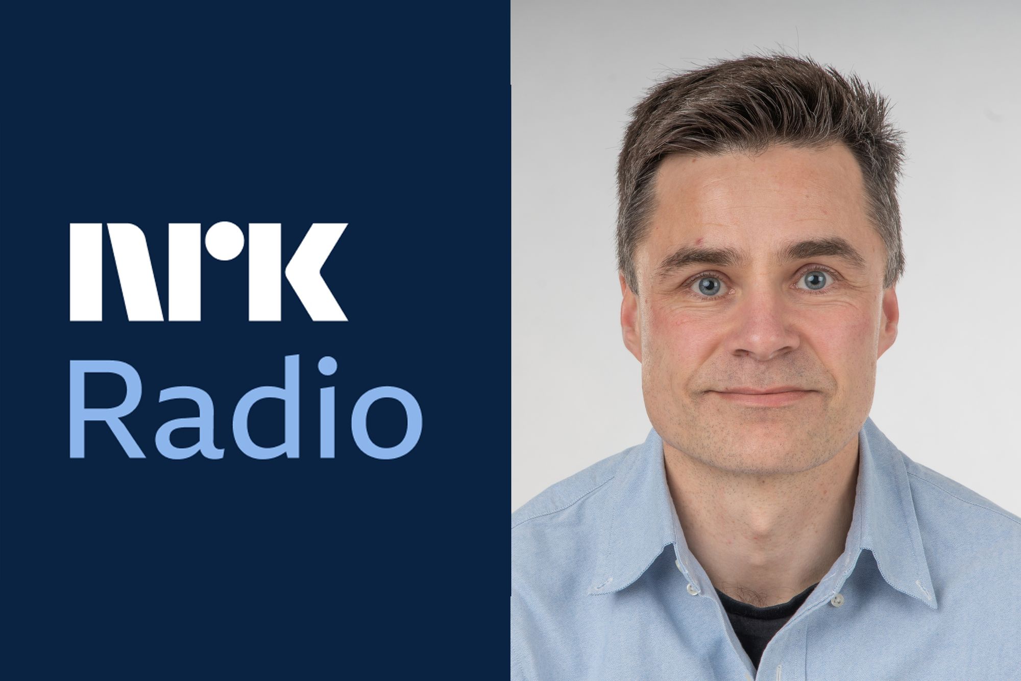
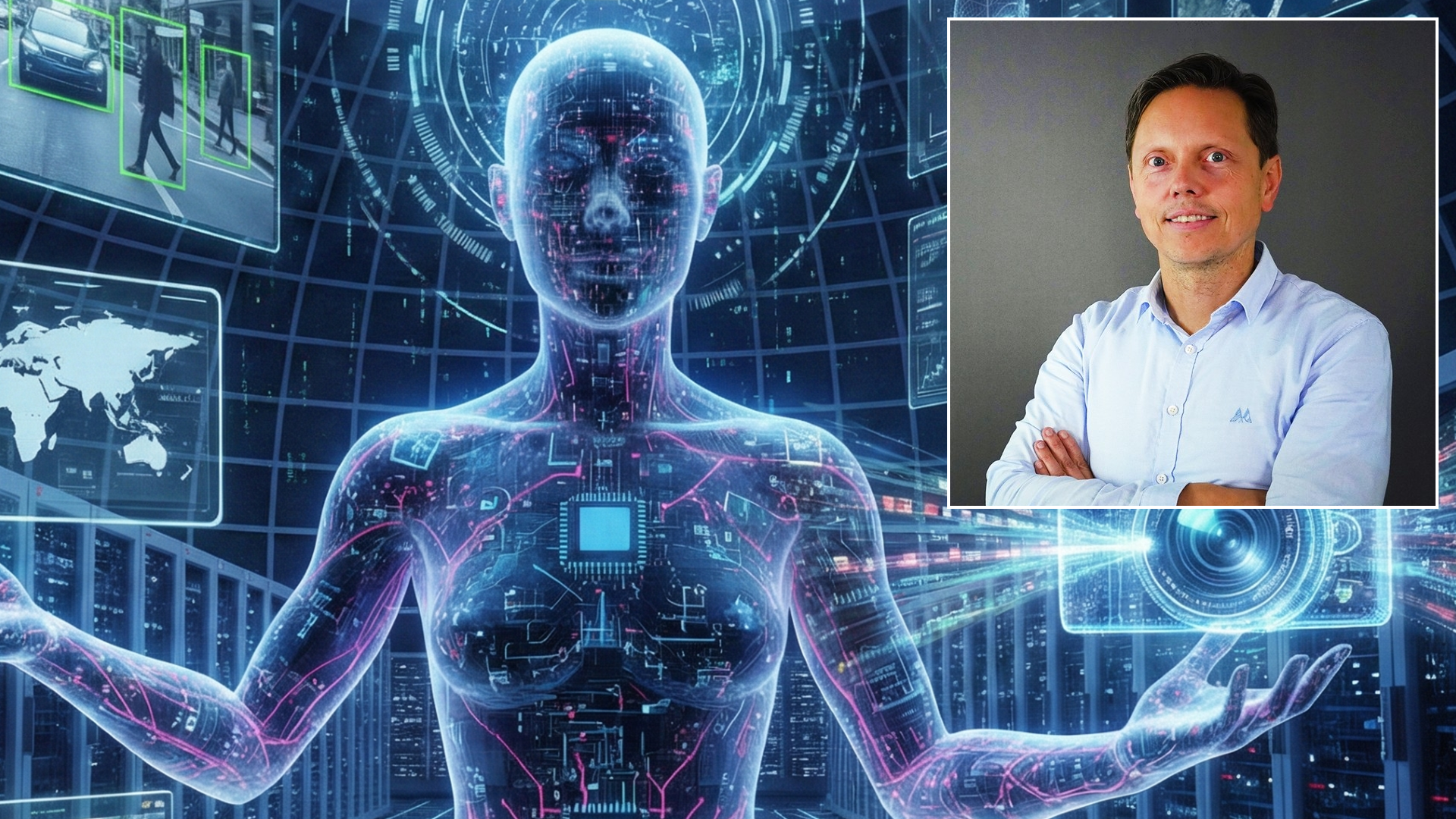
Centre Director Robert Jenssen was interviewed by Norsk rikskringkasting (NRK) about the Japan-Norway AI Innovation Forum in Tokyo, Japan.

En ny AI-løsning utviklet ved UiT kan gjøre det enklere å avdekke feilrapportering og ulovlig fiske. Nå skal Norges Råfisklag teste teknologien i praktisk bruk (News article on kystogfjord.no)
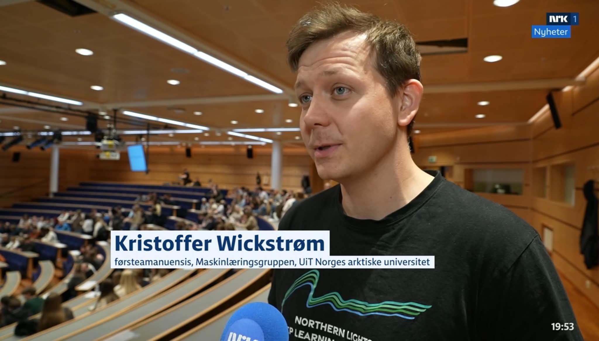
Principal Investigator Kristoffer Wickstrøm was interviewed by Norsk rikskringkasting (NRK) about the Artificial Intelligence Day at UiT- The Arctic University of Norway.
.jpg)
En ny, åpent tilgjengelig KI-modell kan endre hvordan geologer tolker seismikk. Den norske grunnmodellen lover raskere analyser, lavere terskel for innovasjon og nye måter å forstå undergrunnen på.
Centre Director Robert Jenssen was interviewed by Norsk rikskringkasting (NRK) about AI in fisheries and this year's UArctic (University of the Arctic) Congress on the Faroe Islands.
.jpg)
Professor and AI expert Robert Jenssen is attending the Japan-Norway AI Innovation Forum and Japan-Norway Research Symposium, two high-level meetings with Norwegian and Japanese government leaders, business actors and researchers.
Visual Intelligence researchers Solveig Thrun and Kristoffer Wickstrøm took their research out of the lab and to Tromsø city centre as part of Pint of Science 2026.
Subsurface Digital Manager and former Visual Intelligence board member Cathrine Tegnander will serve as the elected leader of Visual Intelligence's board.
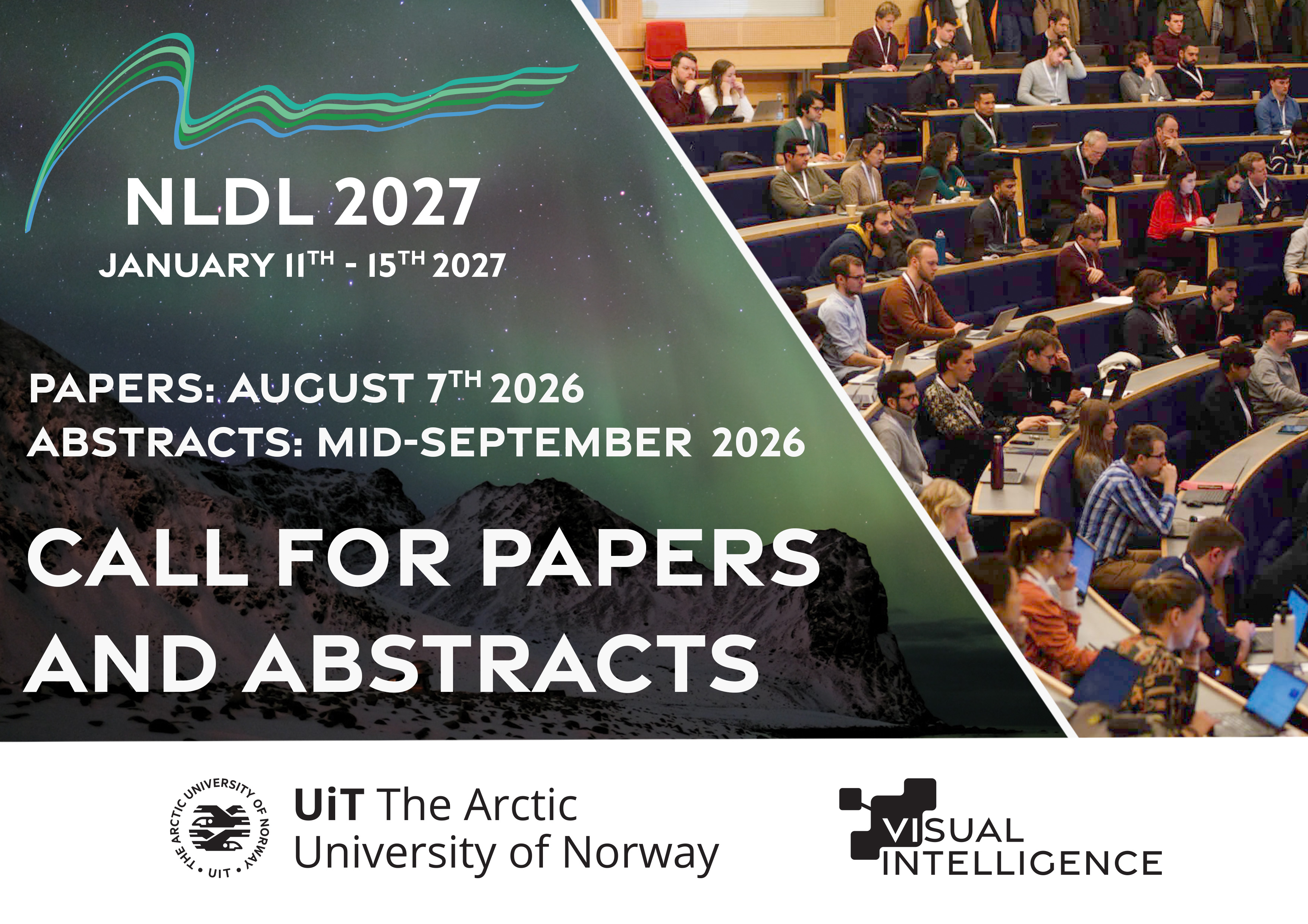
The Call for Papers and Abstracts for the Northern Lights Deep Learning (NLDL) Conference 2027 is officially announced – with submission deadlines on August 7th and Mid-September 2026 respectively.
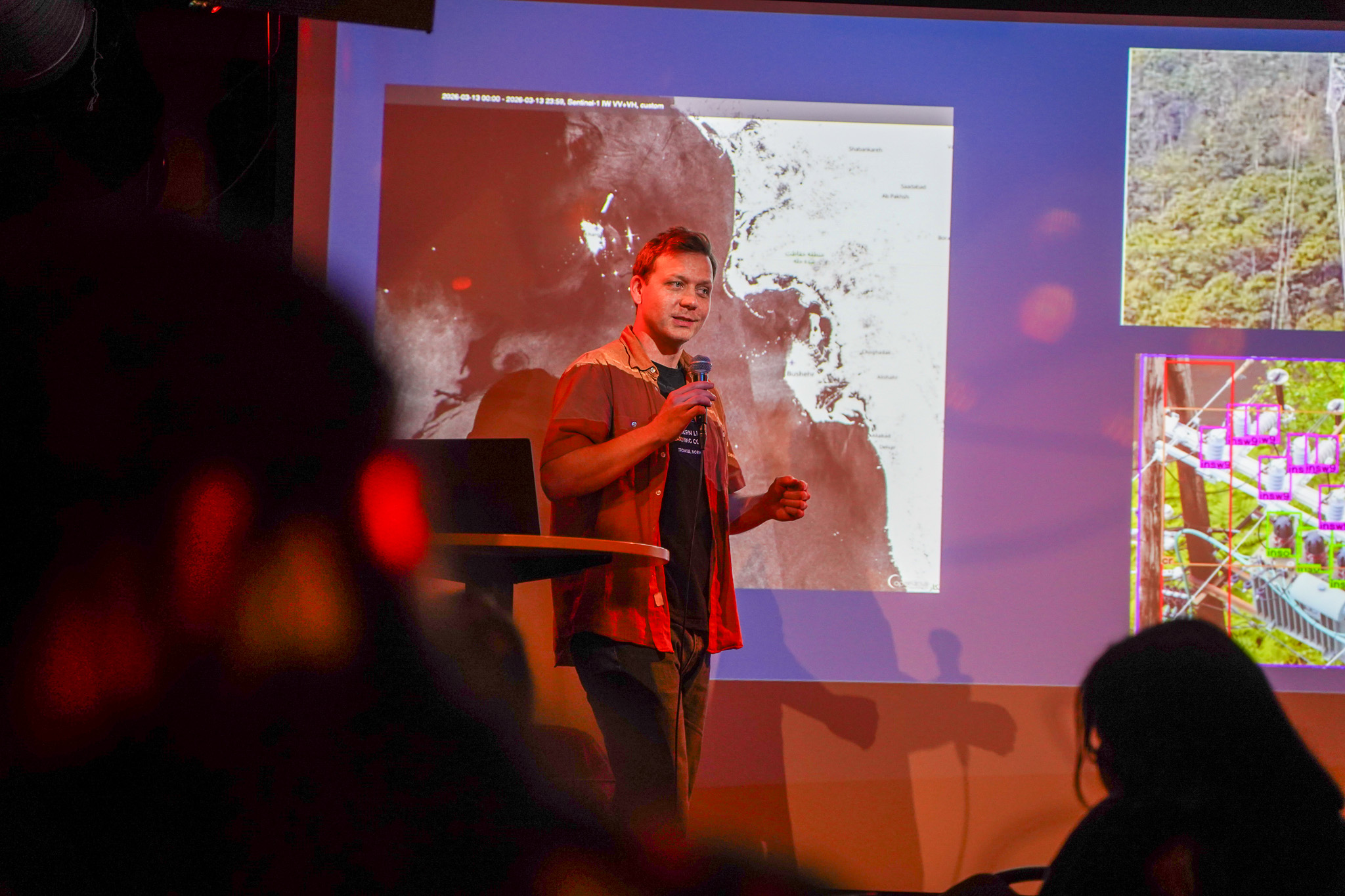
The field of Visual Intelligence is continuously transforming. Chief Research Scientist Arnt-Børre Salberg dives deeper into the current trends in the field of visual intelligence as of early 2026.
Generativ kunstig intelligens er imponerende, men ikke alltid så nyttig til å løse industrielle problemer (Norwegian op-ed in digi.no).
Congratulations to Marit Dagny Kristine Jenssen, who successfully defended her PhD thesis at UiT The Arctic University of Norway on April 10th

The NCS model, a seismic foundation model trained on data from the Norwegian data repository for subsurface data, is now available as an open-source model, allowing anyone to download, utilize, and further develop the model.
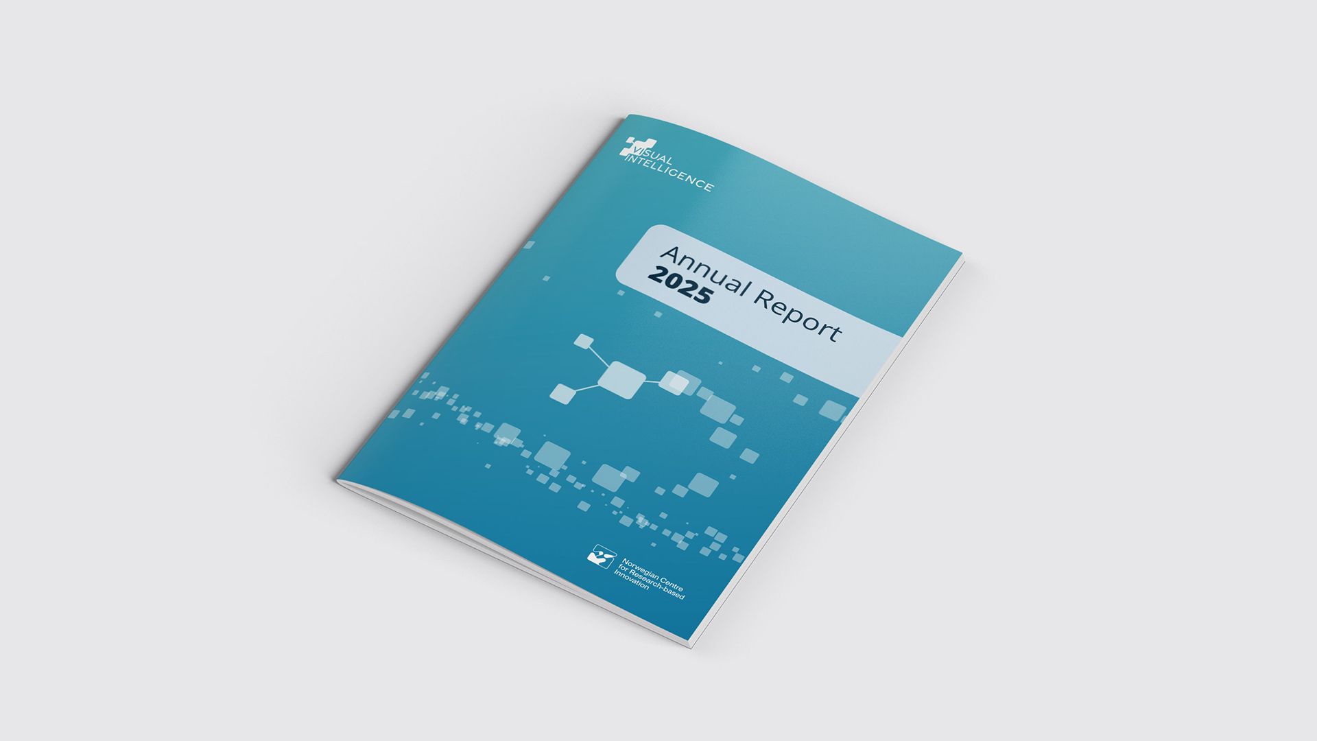
The Visual Intelligence Annual Report 2025, highlighting the centre's progress, activities, achieved innovations, staff, funding, and publications for 2025, is now available to read on our websites.
.jpg)
Congratulations to Centre Director Robert Jenssen, who received UiT's Research and Development Award at the university's annual celebration.
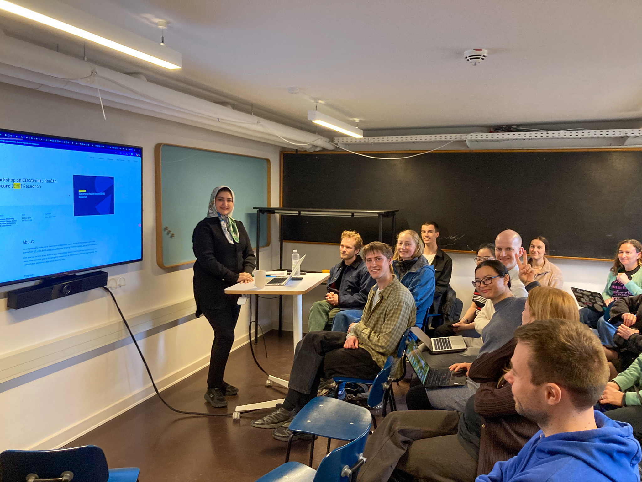
Visual Intelligence researchers contributed to the Pioneer Centre for AI workshop on Electronic Health Records research. The aim was to strengthen ties between the two centres on EHR-related research.